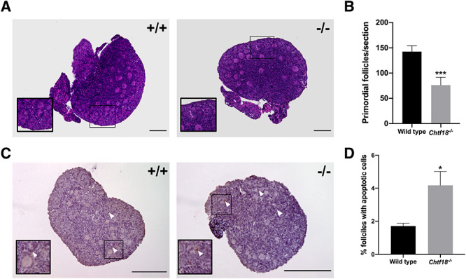
Figure 4.

Chtf18 −/− ovaries have a decreased primordial follicle endowment and increased apoptotic cells at birth. (A) Representative images of paraffin-embedded H&E-stained sections of ovaries from wild-type and Chtf18−/− postnatal day 3 mice. Insets show primordial follicles. (B) Bar graph shows the total numbers of primordial follicles per section as the mean ± SEM; wild-type n = 13 sections (4 mice); Chtf18−/−n = 10 sections (3 mice); and P = 0.0005*** by the Mann-Whitney U test. (C) Representative images of paraffin-embedded sections of wild-type and Chtf18−/− ovaries at postnatal day 3 immunostained with cleaved caspase-3 antibody and counterstained with hematoxylin (apoptotic cells, brown). Arrowheads indicate apoptotic cells in follicles. (D) Bar graph represents the percent ovarian follicles with apoptotic cells as the mean ± SEM; wild-type n = 15 sections (5 mice); Chtf18−/−n = 12 sections (4 mice); and P = 0.0144* by the unpaired t-test. Scale bars, 200 μm.
