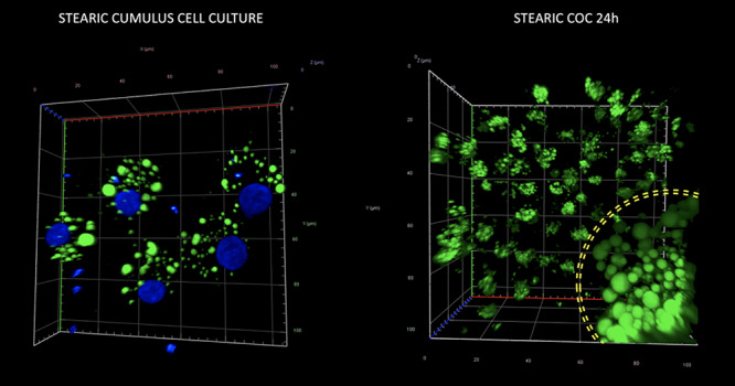
Figure 7.

Morphology and quantity of lipid droplets in cultured porcine cumulus cells (left panel) and a cumulus–oocyte complex (right panel) after supplementation of media with stearic FA. The yellow circle on the right panel corresponds to the zona pellucida and indicates LD population of the oocyte surrounded by several, expanded cumulus cells. Staining with Bodipy 493/503 and DAPI to visualize lipid droplets (green) and chromatin (blue), respectively.
