FIGURE 3.
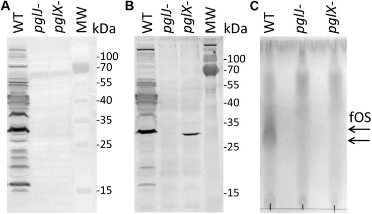
Product analysis of Cf WT, pglX- and pglJ- strains. (A) Western blot of whole-cell lysates with Cff-N-glycan-specific antiserum, and (B) wheat germ agglutinin reactivity of whole cell lysates of the WT, pglJ-, and pglX- strains. (C) Thin-layer chromatography (TLC)-free oligosaccharide (fOS) analysis of WT, pglJ-, and pglX- strains. Molecular weight (MW) markers for the western blots (in kDa) are indicated on the right; arrows indicate the migration of WT fOS on the TLC plate.

