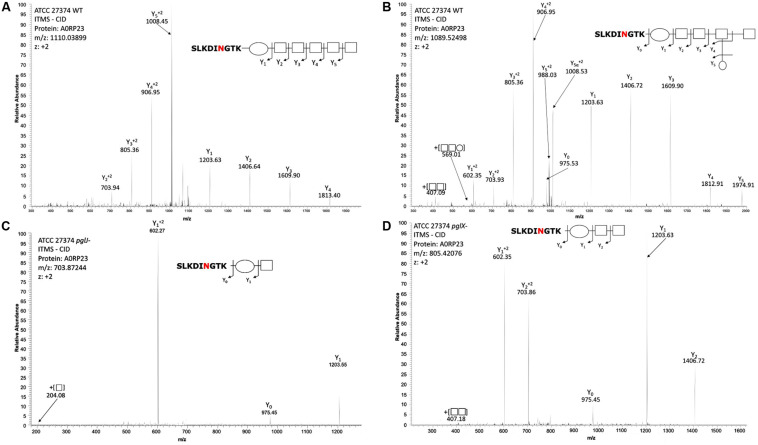
FIGURE 4.
Mass-spectrometric analysis of Cff WT, pglJ-, and pglX- glycopeptides. Fragmentation of characteristic ions obtained by precursor ion scanning of digested Cff lysate samples using liquid chromatography-mass spectrometry. Red lettering indicates possible glycosylation site. Spectra of WT Cff have two glycans: (A) HexNAc5-diNAcBac and (B) HexNAc-[Hex]-HexNAc3-diNAcBac. (C) The peptide from the pglJ- mutant only shows the presence of a mass consistent with HexNAc-diNAcBac. (D) The peptide from the pglX- mutant indicates that it is modified with HexNAc-HexNac-diNAcBac.

