Figure 6.

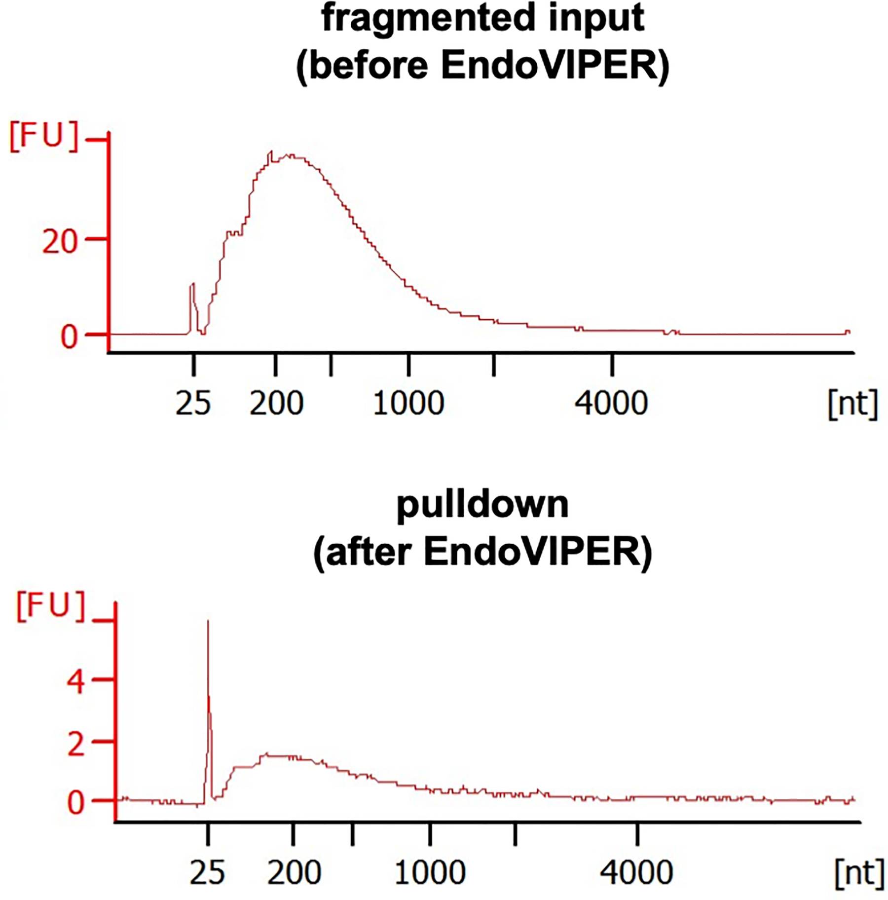
Representative Bioanalyzer electropherograms of fragmented mRNA samples before and EndoVIPER pulldown. Fluorescent units (FU) should be smaller from mRNA after pulldown, but should be roughly the same overall size distribution.

Representative Bioanalyzer electropherograms of fragmented mRNA samples before and EndoVIPER pulldown. Fluorescent units (FU) should be smaller from mRNA after pulldown, but should be roughly the same overall size distribution.