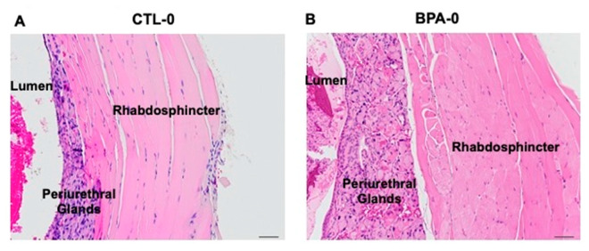
Figure 2.
Post-prostatic urethra between the prostatic and penile urethra. Hematoxylin and eosin stained sections of the urethra wall from adult male mice. (A) CTL-0 male, showing normal lumen and urethra wall: periurethral mucus glands, rhabdosphincter muscle. (B) BPA-0 male showing thickened urethra wall due to glandular hyperplasia and an increase in thickness of the rhabdosphincter (20× magnification).

