
Figure 2.
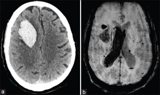
(a) NCCT head revealing large right frontal lobar bleed. (b) SWI sequence of MRI brain showing haemorrhage in right frontal lobe with intraventricular extension and microhaemorrhages in bilateral cerebral hemispheres
Official websites use .gov
A
.gov website belongs to an official
government organization in the United States.
Secure .gov websites use HTTPS
A lock (
) or https:// means you've safely
connected to the .gov website. Share sensitive
information only on official, secure websites.
(a) NCCT head revealing large right frontal lobar bleed. (b) SWI sequence of MRI brain showing haemorrhage in right frontal lobe with intraventricular extension and microhaemorrhages in bilateral cerebral hemispheres