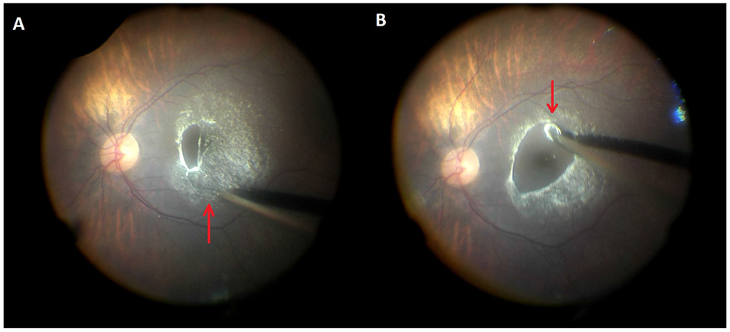
Figure 1.

Careful induction of a posterior vitreous detachment with Flex Loop instrumentation over the macula is performed. In (A), the loop size is made larger (red up arrow) to allow for less tension on the instrument and better visualization whereas in (B), the loop size is smaller (red down arrow) to permit for better tractional force and to separate the hyaloid off the macula.
