
Figure 7. Silencing of Hepatocyte Gramd1b/c in Mice with Hepatosteatosis Prevents TAZ Increase and Suppresses NASH Progression.
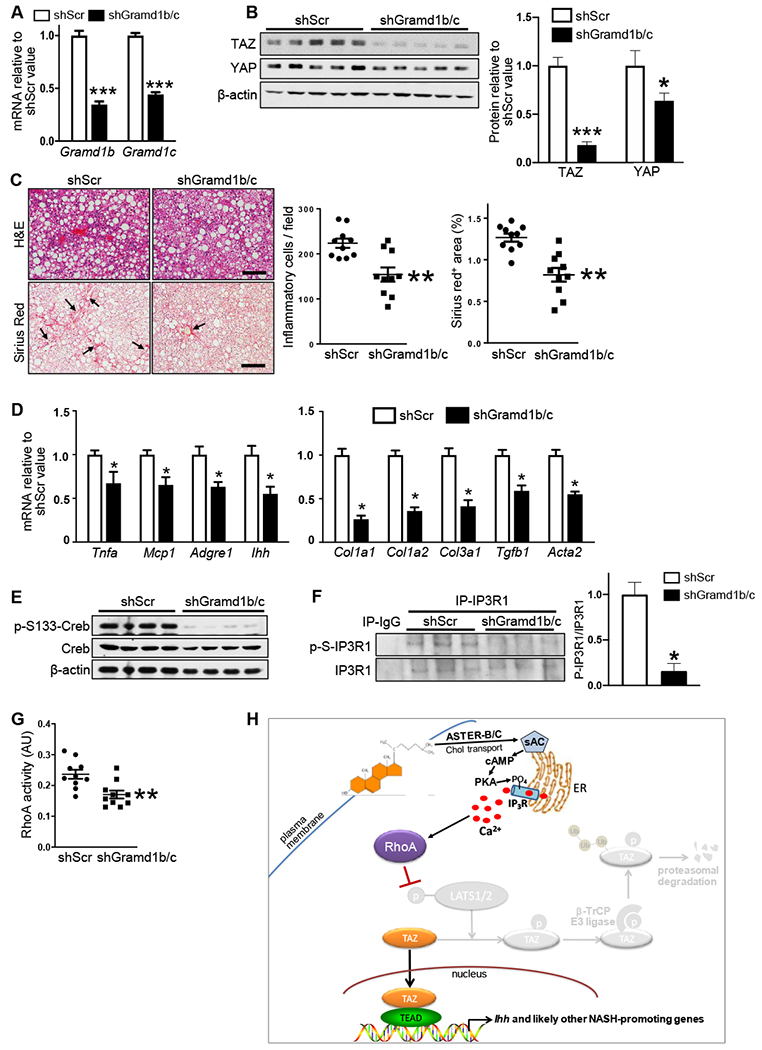
The following parameters were assayed in mice fed the NASH-inducing 1.25%-cholesterol diet for 16 weeks, with AAV8-H1-shGramd1b/c or AAV8-H1-shSrc administered at the 8-week time point (n = 10 mice/group; means ± SEM; *p < 0.05, **p < 0.01, ***p < 0.001):
(A) Liver Gramd1b and Gramd1c mRNAs.
(B) Liver TAZ and YAP immunoblots for 5 mice/group, with quantification.
(C) Liver sections stained for H&E, quantified for inflammatory cells, and stained and quantified for Sirius red (arrows indicate areas of fibrosis). Scale bars, 200 μm.
(D) Liver Tnfa, Mcp1, Adgre1 (F4/80), Ihh, Col1a1, Col1a2, Col3a1, Tgfb, and Acta2 mRNAs.
(E) Liver p-S133- and total Creb Immunoblots.
(F) Phospho-IP3R1 and total IP3R1 immunoblots of liver IP3R1 immunoprecipitates, with quantification.
(G) RhoA activity of liver extracts.
(H) Summary scheme of the hepatocyte cholesterol-TAZ pathway in NASH. In cholesterol-enriched hepatocytes, internalization of plasma membrane cholesterol by ASTER-B/C leads to sAC-mediated increase in cAMP and PKA-dependent activation of IP3R. The resulting increase in Ca2+i activates RhoA, which inhibits LATS1/2-dependent phosphorylation of TAZ. Phosphorylation of TAZ, particularly on S117, blocks β-TrCP-dependent proteasomal degradation of TAZ, resulting in an increase in TAZ-TEAD-induced genes, notably Ihh, which promotes HSC activation and liver fibrosis in NASH (Wang et al., 2016).