Figure 2:

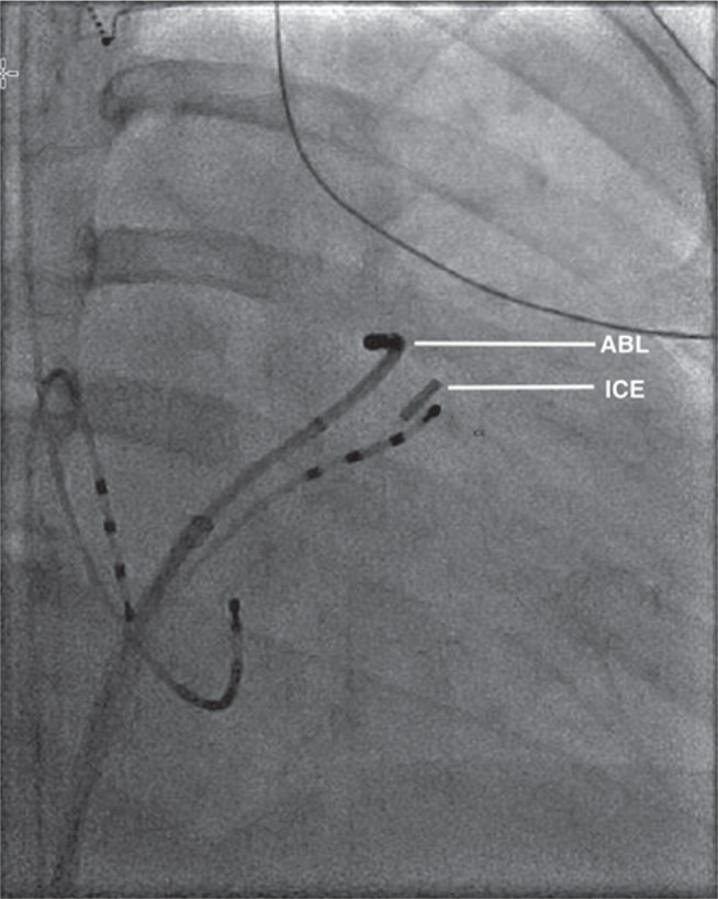
Biplane fluoroscopy image (right anterior oblique: 30 degrees) of the ablation catheter (ABL) positioned across the pulmonary valve, with the ICE catheter positioned in the proximal RVOT.

Biplane fluoroscopy image (right anterior oblique: 30 degrees) of the ablation catheter (ABL) positioned across the pulmonary valve, with the ICE catheter positioned in the proximal RVOT.