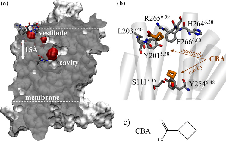
Fig. 2.
OR51E1 conserved motifs form a vestibular-binding site visited by ligands. a Cross section of OR51E1 van der Waals volume (gray). Extra-cellular loops were modeled but are omitted for image clarity. A vestibular-binding site and the orthosteric-binding cavity (red) are detected by a cavity detector. Y2546.48 and V2556.49 at the orthosteric cavity and HRFGTM5 and YGLTM6 forming the vestibule are shown in licorice. b Superpositions of typical positions of CBA (shown in brown) at the cradle of the orthosteric-binding cavity (S1113.36 and Y2546.48) and at the vestibular site. c Chemical structure of CBA

