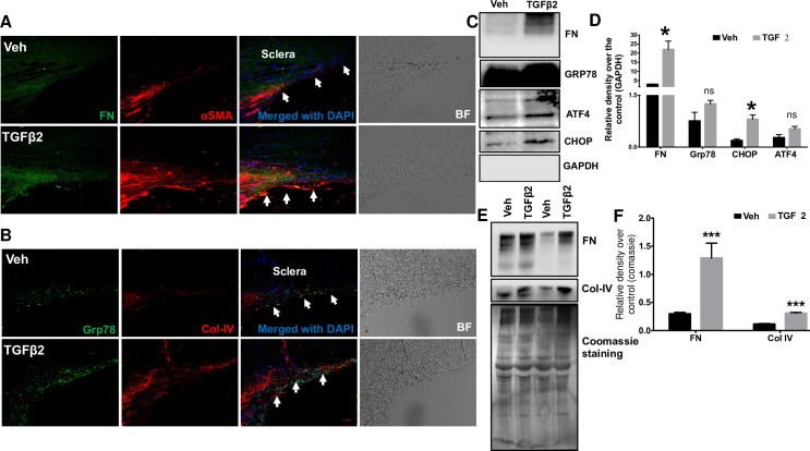
Fig 5. Increased ECM accumulation in the TM of TGFβ2-treated cultured corneoscleral segments.
(A and B) Immunostaining for fibronectin (FN), collagen IV (Col-IV), αSMA and GRP78 in vehicle and TGFβ2 (5ng/ml) treated cultured corneoscleral segments. (n = 4 biological replicates, scale bar is 50μm). Western blot and densitometric analysis for FN, Col-IV (ECM markers), ATF4, CHOP, GRP78 in the TM tissue lysates (C-D) and conditioned medium (E-F) of vehicle and TGFβ2-treated cultured corneoscleral quadrants (n = 4 biological replicates for lysates and n = 8 for the conditioned medium), unpaired t-test, *P<0.05, **P<0.01, ***P<0.001. Arrows indicate the TM region.

