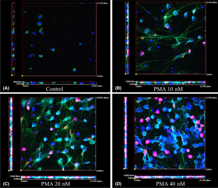
FIGURE 1.

Characterization of PMA induced NETosis by DNA, MPO, CitH3 staining, and visualized by confocal microscopy. Purified neutrophils were incubated on collagen coated coverslips and untreated, A, treated with 10 nM, B, 20 nM, C, or 40 nM, D, of PMA for 3 hours. At the end of treatment, coverslips were fixed with PFA and stained for MPO and CitH3, and mounted with DAPI. Z‐stacks were taken (average of 10 sequential images of 1 μm each) and 3D views were generated with the FV1000 software. (DAPI‐blue, MPO‐green, and CiitH3‐red). Biologic triplicates were performed for each experiment
