FIGURE 4.

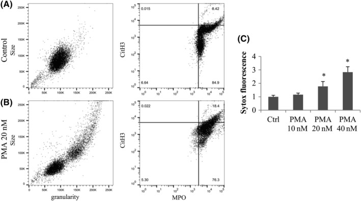
Characterization of PMA induced NETosis by flow cytometry and extracellular DNA assay. Fresh neutrophils were incubated 1 hour with or without PMA, and were fixed and stained with conjugated anti‐CD45 anti‐CD15 antibodies; and anti‐MPO and anti‐CitH3 antibodies followed by secondary antibodies. Cells were gated on the CD45 + CD15+ events and the scatter is shown (size and granularity graph left panel) and MPO in function of CitH3 fluorescence intensity (right panel). A, Untreated neutrophils. B, PMA 20 nM treated neutrophils. C, Attached neutrophils were exposed to 10 to 40 nM of PMA for 3 hours and the extracellular DNA was digested with Mnase, transferred to a new plate and stained with Sytox green. Results are expressed normalized to the control of each experiment and averaged (n = 3 independent experiments). *Statistically different from untreated control
