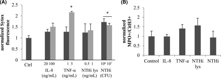
FIGURE 6.

Extracellular DNA detection and flow cytometry analysis of cytokines and NTHi effect on NETosis. A, Treatments (PMA, IL‐8, TNF‐α, NTHi lysates, and live NTHi during 3 hours) were performed and the extracellular DNA was digested with Mnase and transferred to a new plate. Sytox green was then added to stain the DNA. B, Fresh neutrophils were incubated 1 hour in with or without inductor, fixed and stained with conjugated anti‐CD45 anti‐CD15 antibodies; and anti‐myeloperoxidase and anti‐Histone H3 citrullines antibody as well as secondary antibodies. Cells were gated on the CD45 + CD15+ events and results are showing the normalized MPO + CitH3+ population for each treatment. *Statistically different from the untreated control P < .05
