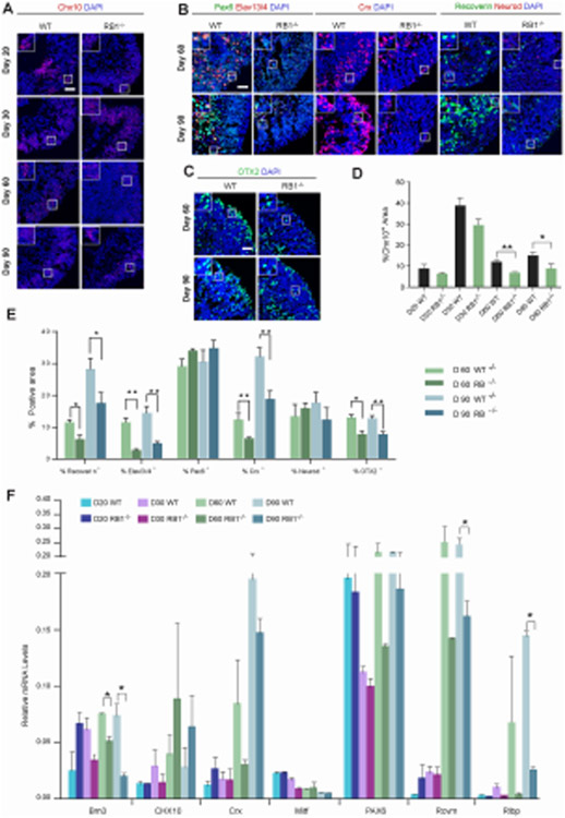
Figure 4. Loss of RB1 reduces photoreceptor, ganglion and bipolar layers.
(A) Representative wild-type and RB1−/− organoids at D20, 30, 60 and 90 sectioned and immunostained against Chx10 and DAPI. (B) Representative wild-type and RB1−/− organoids at D60 and 90 sectioned and immunostained against Elavl3/4 and Pax6; Crx; Recoverin and Neurod; counterstained with DAPI. (C) Representative wild-type and RB1−/− organoids at D60 and 90 sectioned and immunostained against OTX2 and counterstained with DAPI. (D) The graphs show the percentage of Chx10+/DAPI+ area at different time points. (E) The percentage of Elavl3/4+, Pax6+, Crx+, Recoverin+, Neurod+ and OTX2+ cells out of the DAPI+ area at D60 and 90. The results are mean±s.e.m. of 9 organoids from three independent experiments (*P<0.05, **P<0.01, Student’s t-test). (F) Relative mRNA levels of retinal genes measured by qRT-PCR. The results are mean±s.e.m. of 3 organoids from three independent experiments (*P<0.05, **P<0.01, Student’s t-test). Scale bars: 50 μm.

