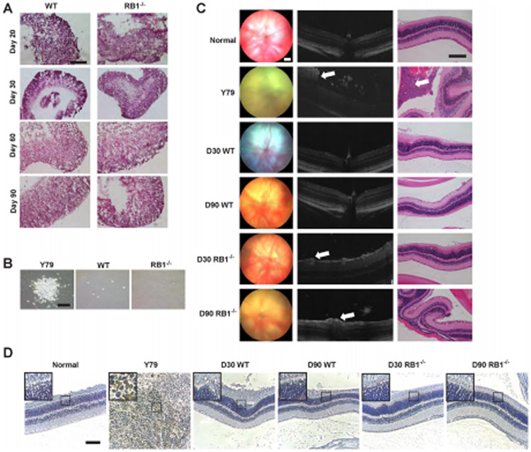
Figure 5. Lack of tumorigenicity in RB1−/− organoids.
(A) Representative images of H&E staining of wild-type and RB1−/− organoids at D20, 30, 60 and 90. (B) Representative brightfield images of colonies using soft agar colony formation assay. (C) The fundus examination (left column), corresponding OCT scans (central column) and histology sections (right column) after intravitreal injection at 12 weeks. Suspicious positive sign (white arrow) of tumor in OCT scan. Verification of tumor (white arrow) in H&E staining. (D) Immunohistochemistry of Ku80, a human-specific nuclear marker. Scale bars: 100 μm.

