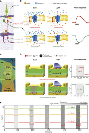
Fig. 1. Retinal and artificial retinal structures.

(A) Profile of a biological retina. (B) Biological working mechanism and photoresponse of OFF bipolar cells [with α-amino-3-hydroxy-5-methyl-4-isoxazolepropionic acid (AMPA)] and ON bipolar cells [with metabotropic glutamate receptor 6 (mGluR6)]. Black bars in the photoresponse of bipolar cells represent the moment of light illumination. (C) Optical image of a retinomorphic device based on a vdW vertical heterostructure. (D) Operating mechanism and photoresponse of the ON- and OFF-photoresponse devices at zero and negative gate voltages, respectively. The positive (negative) ∆Ids corresponds to ON-photoresponse (OFF-photoresponse). Shadow areas correspond to the duration of light illumination. (E) OFF-photoresponse at different bias voltages and light intensities (indicated by shadow areas). OFF-photoresponse of the device remains retained at extremely low bias voltage (10 mV), which allows the operation of low power consumption.
