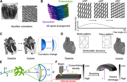
Fig. 1. Design of a physiologically adaptable cardiac patch.

(A) Photograph of the anatomical heart and the fiber structure of the LV visualized by DTI data. (B) Schematic illustration of a short-sectioned LV that illustrates the variation of fiber angle from the epicardium to the endocardium. The orientation (2D mesh pattern projection) of the fiber angles varies continuously with the position across the wall and distribution changes from the apical region to the basal region. (C) Curvature change of cardiac tissue at two different phases (diastole and systole) of the cardiac cycle, which occurs as the heartbeat and pumping blood. (D) CAD design of 3D stretchable architecture on the heart. It provides dynamic stretchability without material deformation or failure when the heart repeatedly contracts and relaxes. (E) Representation of a simplified geometric model of the fibers in the printed object. In the selected region, the angle (α), the length of fiber (L), spatial displacement (D) and the ventricular curvature (κ) are defined with systole (1) and diastole (2) states. (F) Mechanism of the internal stress-induced morphing process. Uneven cross-linking density results in different volume shrinkage after stress relaxation. Photo credit: Haitao Cui, The George Washington University (GWU).
