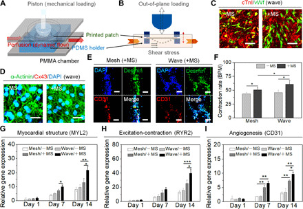
Fig. 4. Biomechanical stimulation and functional maturation of printed cellularized patches.

(A) Schematic illustration of a custom-made bioreactor to apply dual MS for the maturation of engineered cardiac tissue. PMMA, polymethylmethacrylate. (B) Both the out-of-plane loading and fluid shear stress applied to the patches. (C) Immunostaining of cTnI (red) and vWf (green) on the wave-patterned patch under MS condition (+MS) versus nonstimulated control (−MS). Scale bars, 50 μm. (D) Immunostaining of the α-actinin (green) and Cx43 (red) on the wave-patterned patch under MS condition (+MS) versus nonstimulated control (−MS). Scale bars, 20 μm. (E) Cross-sectional immunostaining of the sarcomeric structure (Desmin; green) and vascular CD31 (red) on the patches under MS condition (+MS). Scale bars, 50 μm. (F) The beating rate of hiPSC-CMs on the printed patches under MS condition (+MS) versus nonstimulated control (−MS) on day 14 (means ± SD, n ≥ 6, *P < 0.05). BPM, beats per minute. Relative gene expression of (G) myocardial structure [myosin light chain 2 (MYL2)], (H) excitation-contraction coupling [ryanodine receptor 2 (RYR2)], and (I) angiogenesis (CD31) on the patches under MS condition (+MS) versus nonstimulated control (−MS) on day 1, day 7, and day 14 (means ± SD, n ≥ 9, *P < 0.05, **P < 0.01, and ***P < 0.001).
