
Fig. 5. MTK1 mediates delayed and sustained activation of SAPKs by oxidative stress.
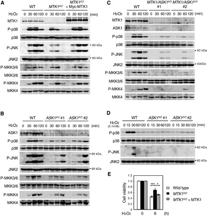
(A) Parental HEK293 cells (WT), MTK1 knock-out cells (MTK1KO), and MTK1KO cells reexpressing Myc-MTK (MTK1KO + Myc-MTK1) were treated with H2O2 (1 mM) for the indicated times. Phosphorylation levels of p38, JNK, MKK3/6, and MKK4 were analyzed by immunoblotting using appropriate phospho-specific antibodies (second, fourth, sixth, and eighth rows). The expression levels of MTK1, p38, JNK, MKK3/6, and MKK4 in cell lysates are also shown (top, third, fifth, seventh, and bottom). (B and D) ASK1 mediates early activation of SAPKs by oxidative stress. HEK293 cells and ASK1-KO cells (clones #1 and #2) were stimulated with H2O2 [1 mM for (B) and 0.1 mM for (D)]. Cell lysates were analyzed by immunoblotting for the expression levels and phosphorylation states of the indicated proteins as in (A). (C) MTK1 and ASK1 are major mediators of SAPK activation by oxidative stress. HEK293 and MTK1/ASK1 double-KO cells (clones #1 and #2) were stimulated with H2O2 (1 mM). The expression levels and phosphorylation states of the indicated proteins were analyzed by immunoblotting. (E) MTK1 promotes H2O2-induced cell death. The indicated cells were treated with H2O2 (0.5 mM) for 6 hours. Cell viability was assessed using the CCK8 assay. Data are means ± SEM (n = 3). *P < 0.05; ***P < 0.01.