
Fig. 4. MD increases following directed microglia interventions.
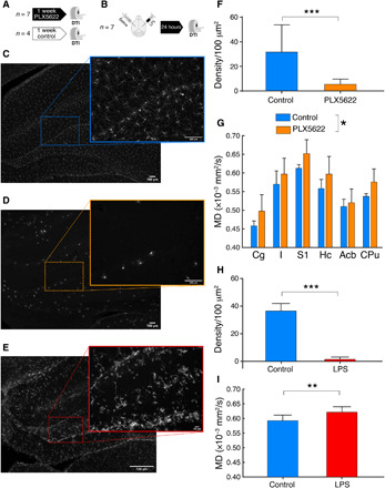
(A and B) Experimental designs for PLX5622 and lipopolysaccharide (LPS), respectively. (C) Representative Iba-1 staining in the hippocampus of a control rat. (D) Representative Iba-1 staining in the hippocampus after 7 days of PLX5622 administration. (E) Representative Iba-1 staining of an LPS-injected hippocampus 24 hours after surgery. (F) Quantification of microglial processes density for control rats and PLX5622 (unpaired t test, t = 7.6, df = 467, P < 0.0001). (G) MD measured in six gray matter ROIs for control rats and rats treated with PLX5622. F(1,8) = 6.3, P = 0.03. (H) Reduction of microglial processes density in the LPS-injected hemisphere versus control (unpaired t test, t = 69.11, df = 237, P < 0.0001). (I) Increase MD in the LPS-injected hemisphere versus control (paired t test, t = 3.7, df = 6, P = 0.009). *P < 0.05, **P < 0.01, and ***P < 0.001. Cg, cingulate cortex; I, insular cortex; S1, primary somatosensory cortex; Hc, hippocampus; Acb, accumbens; CPu, caudate-putamen.