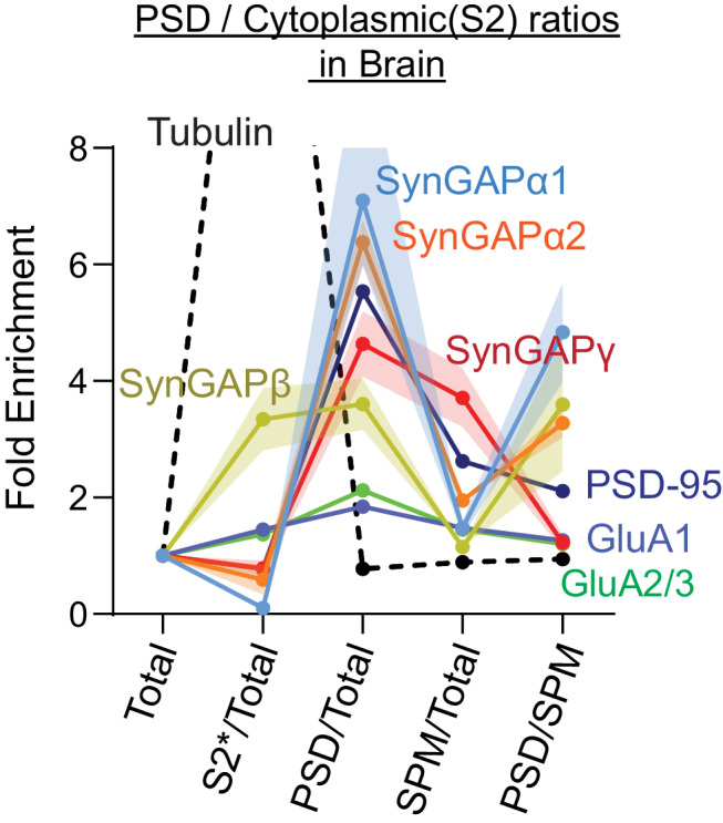
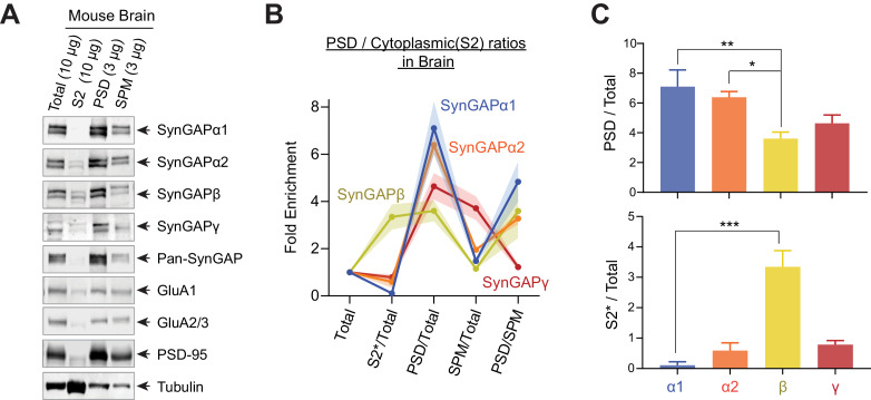
Figure 3. Subcellular localization of various SynGAP isoforms in the brain.
(A) Immunoblot probing endogenous levels of individual SynGAP isoforms and other synaptic proteins in forebrain tissue lysates obtained from adult mice subjected to postsynaptic density fractionation. (B, C) Averaged enrichment of SynGAP isoforms in subcellular fractions in comparison to their levels within the total homogenate fraction, S2 fractions, and PSD fractions. Error bars indicate ± SEM. Kruskal-Wallis test followed by Dunn’s multiple comparison (PSD: H(4) = 15.98; p=0.0011, S2: H(4) = 18.23, p=0.0004, n = 4–7 independent samples for each molecules, Dunn’s multiple comparison ***p<0.001, **p<0.01, *<0.05) was performed.
Figure 3—figure supplement 1. Subcellular localization of various SynGAP isoforms in the brain.


