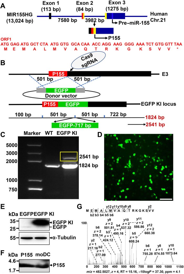
Fig. 1. Discovery of an endogenously expressed micropeptide encoded by MIR155HG.

(A) Schematic representation of P155 translation. Human MIR155HG spans 13,024 bp and has three exons. P155 is translated by ORF1 (indicated by yellow boxes), which comprises the end of exon 2 and the head of exon 3. The nucleotide and amino acid sequences of ORF1 are highlighted in red and the pre–miR-155 is indicated by a bluish color. (B) Schematic representation of P155 EGFP knock-in strategy. The EGFP (without its own ATG) was inserted after the last coding codon (GTT-valine) of P155 by CRISPR/Cas9-mediated homologous recombination in HEK293T cells. The front homologous arm is a 501-bp fragment before the termination codon of P155 sequence and the back homologous arm is a 501-bp fragment starting with the P155 termination codon, E3: exon 3. (C) PCR detection of EGFP knock-in efficiency. Target band is indicated by the yellow box. (D) Fluorescence imaging of P155-EGFP fusion protein expression. (E) Immunoblotting verification of P155-EGFP fusion protein in HEK293T cells. Protein lysate of EGFP plasmid–transfected HEK293T cells served as a negative control. The target band is indicated by black arrowheads, and the EGFP location is visible as a black line. (F) Immunoblotting detection of endogenously expressed P155 in human moDCs with P155-specific antibody pre-enrichment. Chemically synthesized P155 served as a positive control, and the target band is indicated by the black arrowheads. (G) LC-MS verification of the P155 endogenous expression in OCI-LY-1 cells with P155-specific antibody pre-enrichment. Scale bar, 100 μm. Data (D to F) are representative of three independent experiments. Photo credit: Liman Niu (Shanghai Institute of Immunology, Shanghai Jiao Tong University School of Medicine).
