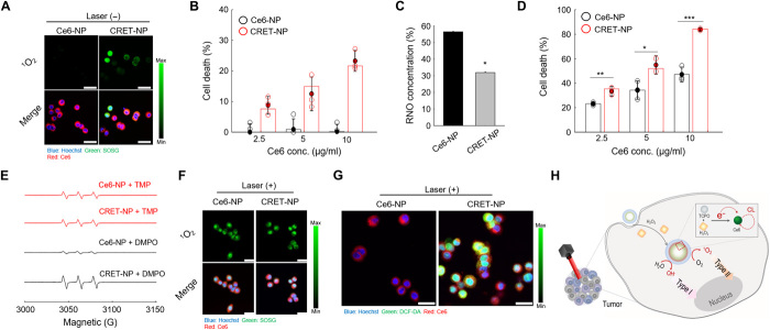
Fig. 5. Characteristics of CRET-NPs as a PDT enhancer.
(A) Confocal microscopy images of 1O2 (green) and NPs (red) in Ce6-NP–treated and CRET-NP–treated HT29 cells. (B) Cytotoxicity of CRET-NPs in HT29 cells. Error bars represent the SD (n = 4). (C) In vitro ROS generation by Ce6-NPs and CRET-NPs under laser irradiation for 30 min. Error bars represent the SD (n = 5). *P < 0.001, analyzed by one-way ANOVA. (D) Cytotoxicity of PDT with Ce6-NPs and CRET-NPs in HT29 cells. Error bars represent the SD (n = 5). *P < 0.05, **P < 0.01, and ***P < 0.001, analyzed by one-way ANOVA. (E) ESR spectra of 1O2 and free radicals from Ce6-NPs and CRET-NPs under laser irradiation for 10 min. (F) Confocal microscopy images of 1O2 (green) and NPs (red) in Ce6-NP–treated and CRET-NP–treated HT29 cells under laser irradiation. (G) Confocal microscopy images of ROS (green) and NPs (red) in Ce6-NP–treated and CRET-NP–treated HT29 cells under laser irradiation. (H) Schematic illustration of ROS quantum yield enhancement by CRET-NPs. Scale bars, 25 μm (A, F, and G).

