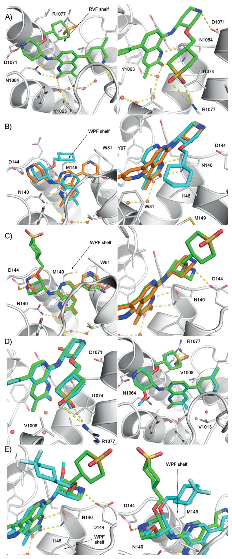
Figure 1.
A) 2 views of the X-ray structure of 3 bound to ATAD2 (PDB 5a83). B) Superimposed X-ray structures of BRD4 BD1 bound to 1 (orange, PDB 5a5s) and 2 (cyan, PDB 5a85). C) Superimposed X-ray structures of BRD4 BD1 bound to 1 (orange, PDB 5a5s) and 4 (green, PDB 5lj2) showing the di-axial conformation of the piperidine ring. D) ATAD2 crystallographic binding modes of 16 (cyan, PDB 5lj0) and 3 (green, PDB 5a83). E) Binding modes in BRD4 BD1 of 16 (cyan, PDB 5lj1) and 4 (green, PDB 5lj2). For refinement statistics see Table S3; for density maps Figure S6, Supporting Information.

