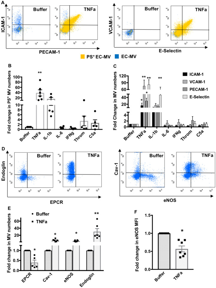
Figure 2.
In vitro inflammatory stimulation alters endothelial MV numbers and cargos. (A) Representative flow cytometry dot plots demonstrate increased ICAM-1, PECAM-1, VCAM-1, E-selectin, and PS positive MVs generated from HUVECs treated with TNFα. (B) PS+ EC-MV generation is significantly increased by TNFα treatment (n = 5–6 experiments). (C) TNFα significantly increased ICAM-1+, VCAM-1+, PECAM-1+, and E-selectin+ EC-MV generation, while IL-1β only significantly increased E-selectin+ EC-MV generation from HUVECs (n = 5–10 experiments). **P < 0.01 between buffer and individual inflammatory mediators by one-way ANOVA analysis and Dunnett’s multiple comparison tests. (D and E) Flow cytometry analyses of TNFα-induced EC-MVs containing EPCR+, Cav-1+, eNOS+, and endoglin+ cargos. *P < 0.05, **P < 0.01 between buffer and TNFα by Student’s t-test, n = 5–6 experiments. (F) eNOS expression (median fluorescence intensity) is significantly reduced in EC-MVs induced by TNFα. *P < 0.05 by Student’s t-test, n = 7 experiments. All data are shown as mean ± SEM.

