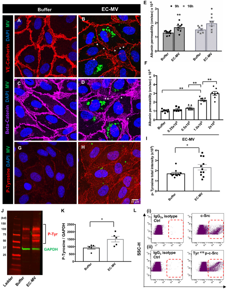
Figure 3.
EC-MVs increase endothelial permeability and induce phosphorylation of proteins in recipient cells. (A–D) Representative confocal images showing discontinuous adherens junctions in HUVEC monolayers after EC-MV treatment. VE-cadherin (A and B) staining in buffer-treated and MV-treated ECs; (C and D) beta-catenin staining in HUVECs with and without EC-MVs (green dots indicate MVs, arrowheads point to discontinuous junction lining). (E) Albumin permeability through endothelial monolayers is increased after treatment with EC-MVs at 9 and 16 h. *P < 0.05, **P < 0.01 between buffer and EC-MV at 9 h and 16 h, respectively by Student’s t-test, n = 7–8 experiments. (F) Albumin permeability through endothelial monolayers is increased after treatment with increasing concentration of EC-MVs. **P < 0.01 by one-way ANOVA with Tukey’s multiple comparison tests, n = 6–7 experiments. (G–I) Representative confocal images showing phospho-tyrosine level is remarkably increased after EC-MV treatment. Scale bar = 10µm and applies to all images. *P < 0.05 by Student’s t-test, n = 11 different confocal z stacks from multiple slides. (J and K) Western blot analysis showing phospho-tyrosine level is significantly increased after EC-MV treatment. *P < 0.05 between control and EC-MV treated cells by Student’s t-test, n = 5 lysates. All data are shown as mean ± SEM. (L) Representative flow cytometry dot plots show the presence of c-Src kinase and Tyr416 phosphorylated c-Src on EC-MVs.

