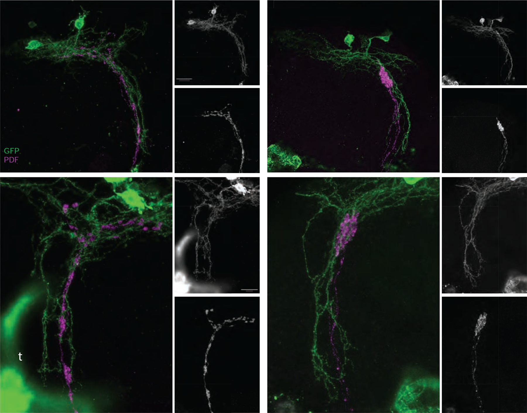
Figure 4. The expression of Unc5 causes significant changes in the anatomical relationship between the neurites of the DN1ps and the dorsal projections of the s-LNvs.

(A-C) Confocal reconstruction of s-LNv dorsal projections and the neurites of the DN1p clock neurons in the dorsal protocerebrum of a ;Pdf-Gal4/LexAop-mCD8:GFP;Clk4.1LexA/+ brain. (A) Brains were immuno-labelled for GFP (green) and PDF (magenta) and imaged through the posterior surface of the brain. Small panels display single gray scale projections of GFP (B) and PDF expression (C). The medial (m), lateral (l) and ventral (v) extensions of the DN1p neurons are indicated in B and the medial (m) and lateral (l) extensions of the s-LNv dorsal termini are indicated in C. (D-F) Confocal reconstruction of s-LNv dorsal projections and the neurites of the DN1p clock neurons in the dorsal protocerebrum of a ;Pdf-Gal4/LexAopmCD8:GFP;Clk4.1LexA/UAS-Unc5 brain immunolabeled and imaged as described for A-C. The major extensions of the DN1ps are intact, yet the medial and lateral extensions of the s-LNvs are completely absent. Merged GFP and PDF signals are shown in D. Small panels display single gray scale reconstructions of GFP (E, labels as for B) and PDF expression (C). (G-H) High magnification confocal reconstruction of the dorsal projections of normal s-LNvs (magenta) and their relationship with the ventral projection of the DN1ps. The genotype is the same as in A and panels organized and labeled as for A-C. High levels of GFP are expressed in the tracheae (t in panel G). (J-L) High magnification projection of an Unc5-expressing s-LNv dorsal projection and neighboring DN1p neurons. The organization of the DN1p ventral extension is unchanged by the truncated s-LNvs. Panels organized and labeled as for D-F. Scale bars = 25 μm for A-F and 15 μm for G-L.
