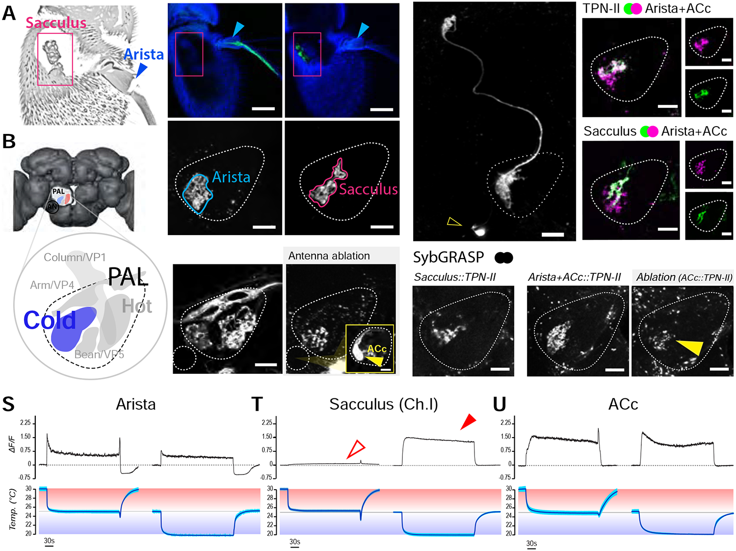
FIGURE 3 |. Three distinct populations of peripheral cold sensing neurons drive the activity of TPN-IIs.

(A) Confocal micrograph of the fly antenna showing the location of the sacculus (pink box) and arista (blue arrow). (B) 3D model of the fly brain showing the location of the posterior antennal lobe (PAL) and PAL glomeruli (inset; the glomerulus innervated by cold cells is shown in blue, additional glomeruli are annotated with standard nomenclature). (C-H) Selective drivers identify distinct sensory neuron populations targeting the cold glomerulus. (C,D) A selective driver for arista cold cells labels (C) cell bodies in the arista and (D) their PAL termini. (E,F) A selective driver for cold cells of the sacculus labels (E) cell bodies innervating chamber I of the sacculus and (F) their termini in the PAL. (C,E are confocal micrographs of the whole antenna; blue: cuticle autofluorescence, green: GFP, scalebars 50μm; D,F are single 2-photon slices; scalebars 10μm). (G,H) Antenna ablation demonstrates the existence of an unusual “internal” cold receptor also innervating the PAL. (G) IR25a-Gal4>UAS-GFP labels many of the antennal sensory neurons innervating PAL glomeruli. (H) A week following antennal resection, all antennal afferents have degenerated, revealing Anterior Cold cell (ACc) termini in the PAL. The fluorescent signal can be traced to one/two cell bodies located on the edge of the antennal nerve (AN, inset –scalebar 10μm; see also Figure S2). (I-O) Extensive overlap between TPN-II dendrites with both arista/ACc and sacculus termini in the PAL. (I) A selective split-Gal4 driver reveals TPN-II’s anatomy (2-photon z-stack). (J-O) 2-color 2-photon micrograph illustrating spatial overlap between (J-L) TPN-II dendrites (green) and Arista/ACc termini (magenta, single z slice), and between (M-O) sacculus (green) and arista/ACc termini (magenta). (P-R) Synaptobrevin GRASP confirms synaptic connectivity between TPN-IIs and (P) sacculus, (Q) arista/ACc; the fact that the syb:GRASP signal in Q persists a week post-antenna ablation (R) shows ACc also connect to TPN-IIs. (S-U) 2-photon Ca2+ imaging shows sacculus neurons exclusively respond in the cold range. Response profiles of (S) arista, (T) chamber I sacculus, and (U) ACc neuron termini in response to cooling steps in the hot (bottom, above 25°C, red) or cold (bottom, below 25°C, blue) range. (S) Arista neurons show both a transient peak in response to cooling and a persistent Ca2+ elevation in both conditions. (T) Sacculus cold cells only respond when the temperature drops below 25°C (arrows). (U) ACc neurons show persistent signals both above and below 25°C. S-U show averages of 4 animals ±SD. In all PAL panels, scale bars are 10μm.
