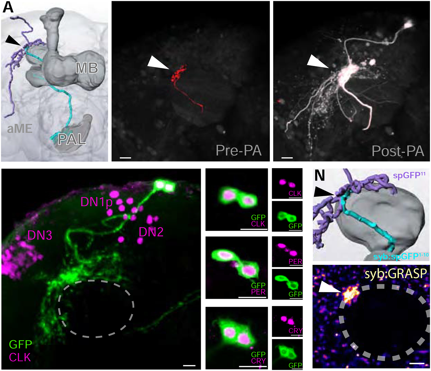
FIGURE 4 |. TPN-IIs target the DN1a group of dorsal neurons, part of the circadian clock network.

(A) 3D reconstruction of the TPN-II (blue)-DN1a (purple) connection, at the edge of the mushroom body (MB) Calyx. (B-C) Photolabeling with PA-GFP reveals DN1as are candidate targets for TPN-IIs. For this experiment, (B, pre-photoactivation) TPN-II termini are targeted by independent labeling with TdTomato, while PA-GFP is expressed broadly. Following targeted photoactivation at 720 nm, (C, white) PA-GFP diffuses to label TPN-II targets. (D-M) DN1as are identifiable by anatomy, and because they express a combination of molecular clock components. Here, (D) DN1as were labeled by GFP expression (green, under the control of a selective driver) and immunostained using an anti-Clock antibody (CLK, purple; confocal z-stack of a whole-mount fly brain, note that CLK also labels DN1p, DN2, and DN3). (E-G) Enlargement of DN1as shown in D. (H-J) DN1as also express Period (anti-PER, purple) and (K-M) Cryptochrome (anti-CRY, purple; in all panels scalebar =10μm). (N-O) Syb:GRASP demonstrates monosynaptic connectivity between TPN-IIs and DN1as. (N) Enlargement of a 3D reconstruction showing predicted point of synaptic contact. (O) Synaptic GFP reconstitution is observed between TPN-II and DN1a neurons at the edge of the mushroom body calyx (pseudocolored 2-photon stack).
