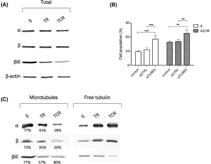
FIGURE 5.

Decreased tubulin βIII expression is associated with decreased polymerized tubulin fraction and increased S and G2/M phase fractions. The protein expression of total α and β tubulin and isoform βIII was studied in total cell lysates (A) or after purification of tubulin fractions (C). (A) Western blot of tubulins α, β, and βIII shows that while total α and β tubulin expression are unchanged, βIII protein expression is decreased in T‐DM1‐resistant cells. The density of each band was normalized to actin. (B) Downregulation of βIII tubulin in the parental cell line leads to increased S and G2/M populations, 48 hour after transfection by siRNA. (C) Tubulin purification was performed to separate the polymerized (microtubules) and soluble (free) tubulin fractions. The percentage values correspond to the amount of polymerized tubulin in each cell line, for each tubulin type. The percentage of total α and β tubulin in microtubules is decreased in resistant cell lines compared to the parental one. The percentage of βIII tubulin in microtubules is decreased in the TR cell line. Even though the percentage of βIII tubulin in microtubules is unchanged in TCR cells compared to parental cells, the density of the bands indicates a higher amount of βIII tubulin in parental than resistant cells in the microtubule fraction
