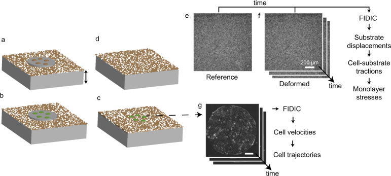
Fig. 1.
Experimental flowchart to measure cell velocities, cell-substrate tractions, and monolayer stresses. (a) A PDMS mask was placed on a polyacrylamide substrate embedded with fluorescent particles (see Methods), and collagen I was adhered to the substrate at locations of the holes in the mask. (b) Cells (green) were seeded to form cellular islands of desired diameter. (c) The PDMS mask was removed, and time-lapse imaging of a confluent cell island and the fluorescent particles was performed simultaneously. (d) The cell island was removed using 0.05% trypsin, which allowed the substrate to recover to a stress-free reference state used for computing cell-substrate tractions. (e,f) The reference image was correlated with the time-lapse images of the deformed substrate to obtain the substrate displacements. (g) Consecutive phase contrast images of the cell island were correlated to compute cell velocities.

