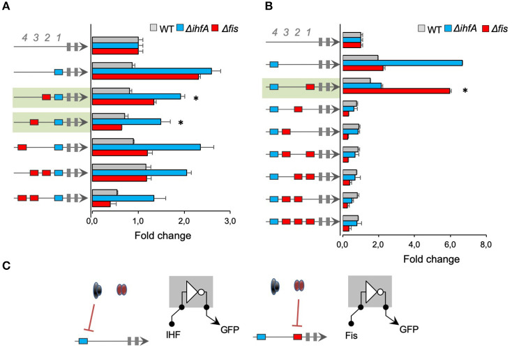
Figure 4.
The activity of promoters with combined IHF- and Fis-binding sites. The architecture of the synthetic promoter is shown on the left (blue boxes represent IHF-BS and red boxes represent Fis-BS). Promoter activities are shown in bars and normalized based on the activity of the reference promoter (i.e., a promoter with 4 neutral sequences). Promoter variants were analyzed in wild type (gray bars) Δfis (red bars), and ΔihfA (blue bars) mutant strains of E. coli. (A) Characterization of promoters with a single IHF-BS fixed at position 1 (−61) and varying Fis-BS. Promoters shaded in green present reduced activity in the ihfA and fis mutant strains, compared to IHF-BS at the 1st position. Statistical differences between synthetic promoters and their control (PNNNI) are highlighted by (*) as analyzed using Student's t-test with p < 0.05. (B) Characterization of promoters with a single IHF-BS fixed position 4 (−121) and varying Fis-BS. Promoter shaded in green displayed increased activity in the E. coli Δfis strain, while it showed no activity in the wild type and ΔihfA strains. Statistical differences between this promoter in Δfis condition and wild type and ΔihfA are highlighted by (*) as analyzed using Student's t-test with p < 0.05. (C) Summary of most significant changes in promoter architecture leading to changes in promoter logic. Statistical differences between synthetic promoters and their control are highlighted by (*) as analyzed using Student's t-test with p < 0.05.

