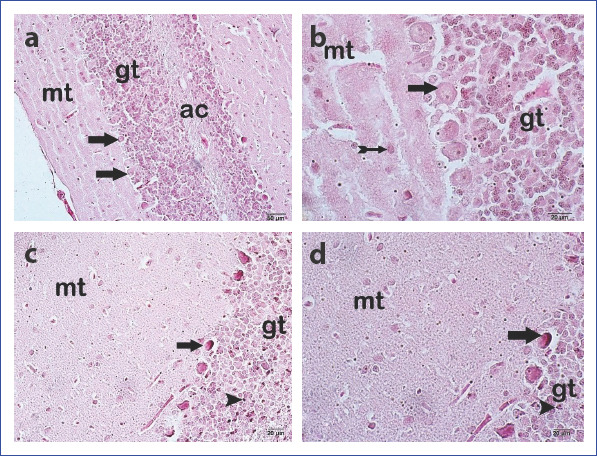
Figure 2 (a, d).

Appearance of the cerebellum under light microscope. Control Group: (a) Purkinje cell (arrow) 200× (b) Purkinje cell (arrow) basket cell (arrow with tail) 400×. Electromagnetic field group (d) Degenerative Purkinje cells with pycnotic nuclei (arrow) and granular cells with pycnotic nuclei (arrowhead) 200×. (e) Degenerative Purkinje cells with pycnotic nuclei (arrow) and granular cells with pycnotic nuclei (arrowhead). Mt, molecular layer; Gt, granular layer; Ac, white matter (H&E).
