. 2020 Jun 25;13(2):183–186. doi: 10.3400/avd.cr.19-00104

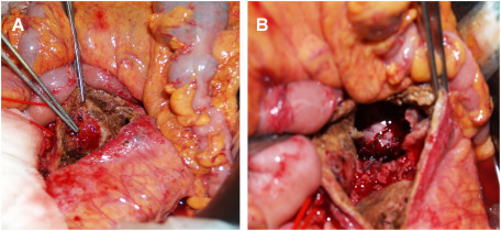
Fig. 2 Intraoperative pictures during the staged laparotomy. (A) Two 3-mm wide fistulas between the anterior wall of the abdominal aortic aneurysm and the third part of the duodenum are identified inside the aneurysm. (B) A deployed stent graft is seen inside the aneurysm, which is isolated and uncontaminated with intestinal fluid.

Fig. 2 Intraoperative pictures during the staged laparotomy. (A) Two 3-mm wide fistulas between the anterior wall of the abdominal aortic aneurysm and the third part of the duodenum are identified inside the aneurysm. (B) A deployed stent graft is seen inside the aneurysm, which is isolated and uncontaminated with intestinal fluid.