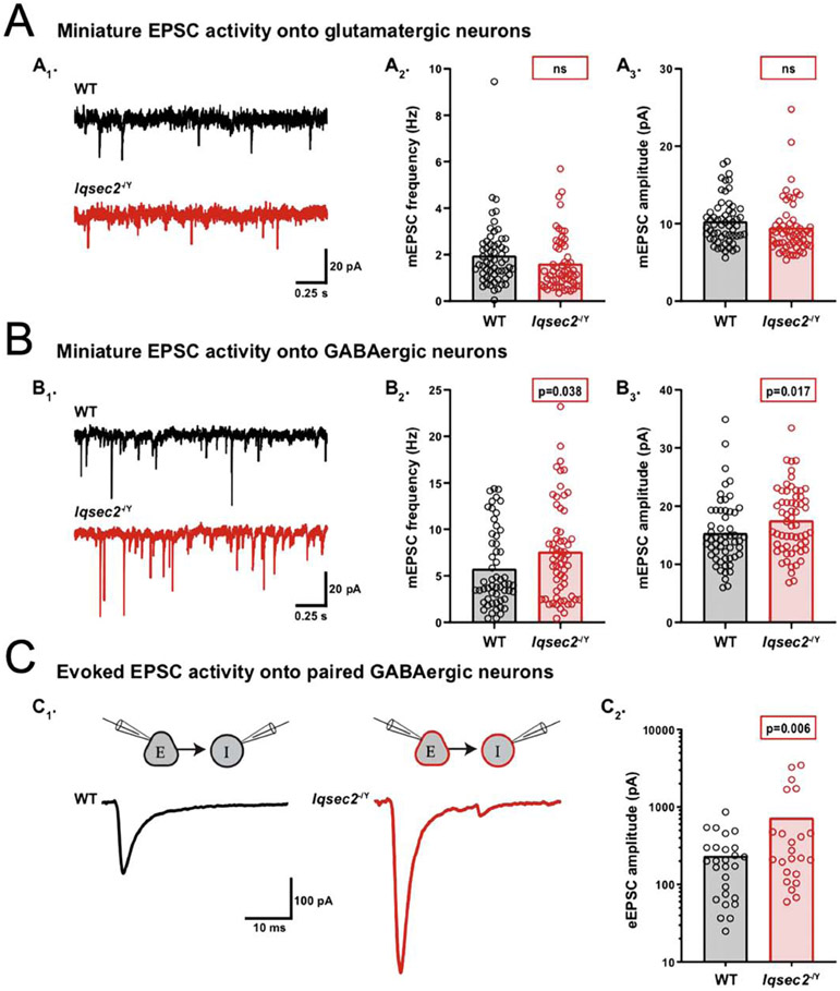
Figure 4: IQSEC2 loss increases excitatory drive onto GABAergic neurons.
A-C. Using whole-cell, voltage-clamp electrophysiology, spontaneous miniature and evoked activity was recorded from glutamatergic and GABAergic hippocampal neurons derived from wildtype (WT) and Iqsec2−/Y mice at days 12–15 in vitro. Example traces of miniature EPSC activity onto wildtype (WT) (black) and Iqsec2−/Y (red) glutamatergic neurons (A1) and GABAergic neurons (B1). Bar graph with overlaid dot plots show that the mEPSC frequency and amplitude onto glutamatergic neurons were both unaltered (A2 and A3), whereas onto GABAergic neurons they were both increased (B2 and B3), by Iqsec2 loss. C1. Example traces of evoked EPSCs onto paired wildtype (WT) (black) and Iqsec2−/Y (red) GABAergic neurons. C2. Bar graphs with overlaid dot plots show that the eEPSC amplitude onto GABAergic neurons was increased by loss of Iqsec2. For all graphs, the bars indicate the mean of the data set and each dot represents the mean response from one neuron. All p-values were calculated using Generalized Estimating Equations, and significant p-values are indicated in red boxes for each graph (ns indicates p>0.05).

