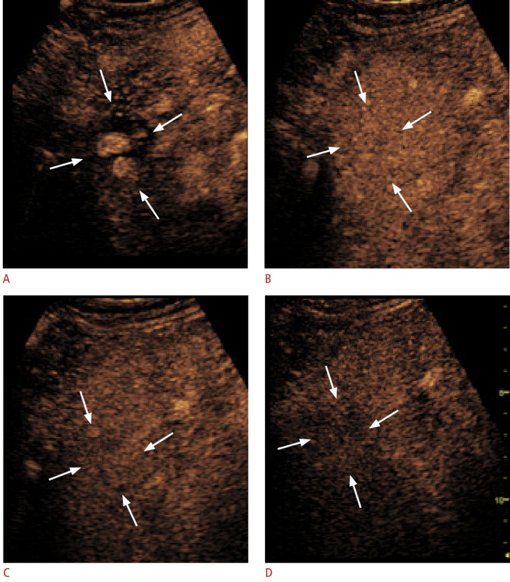
Fig. 2. Nodule-in-nodule appearance.

The patient is a 69-year-old man with alcoholic cirrhosis and an LR-4 lesion on magnetic resonance imaging. A. Contrast-enhanced ultrasound shows a 32-mm nodule with a nodule-in-nodule appearance in the arterial phase (arrows). B-D. The entire nodule (arrows) displays isoenhancement in the portal venous phase at 1 minute (B) and 2 minutes (C) and shows late and mild washout at 3 minutes (D). With regard to appearance, this is typical of a well-differentiated hepatocellular carcinoma focus developed in a dysplastic nodule.
