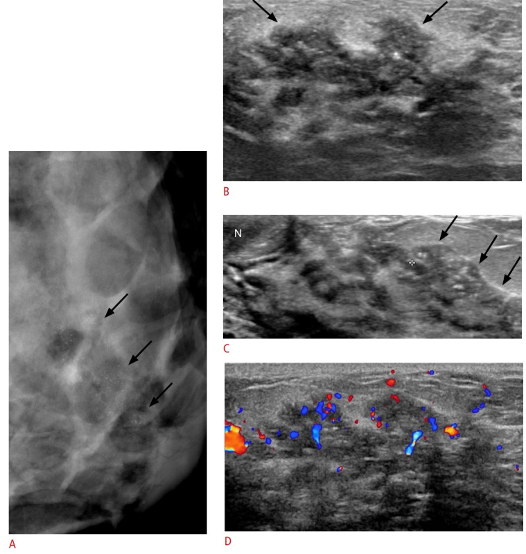
Fig. 12. Pregnancy-associated breast cancer in a 42-year-old pregnant woman (intrauterine pregnancy 35 weeks) presenting with bloody nipple discharge.

A. Mediolateral oblique mammogram shows segmental distributed, fine pleomorphic microcalcifications (arrows). B, C. Ultrasonography shows an irregular ductal change with intraductal calcifications (arrows). D. Color Doppler study shows increased vascularity within the lesion. The patient underwent left breast conserving surgery and histologic findings revealed invasive carcinoma with ductal carcinoma in situ.
