
Figure 2.
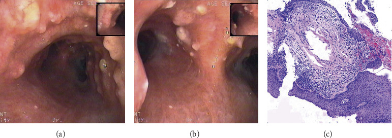
Case 4 (a) bronchoscopic examination: multiple nodular tuberositas of the lower trachea, the surface is uneven and pebble-like, part fuse into pieces. (b) Bronchoscopic examination: multiple nodular tuberositas of the tracheal carina and left and right main bronchus and some surfaces are covered with yellow pus. (c) Hematoxylin and eosin (H&E) stain at 100x magnification: pathological results of tracheal nodules, partial squamous cells on the bronchial mucosa epithelium, a large number of lymphocytes, plasma cells, and partial ossification.