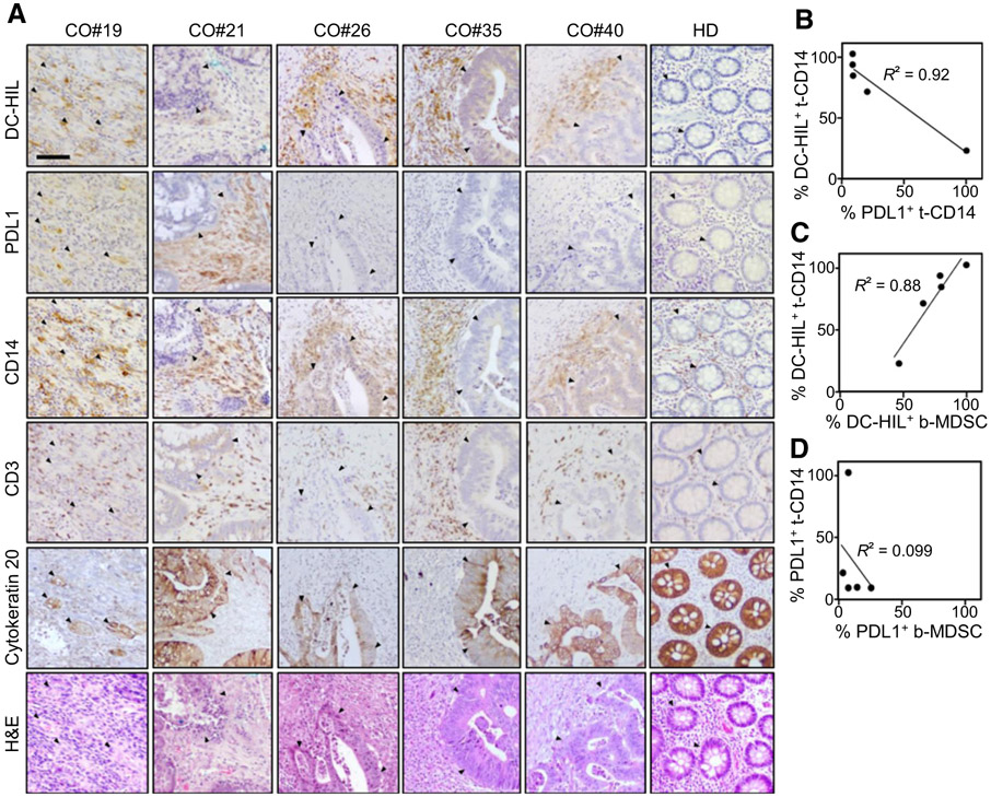
Figure 4.
DC-HIL and PDL1 expression in colorectal tissues of patients with cancer. A, Serial sections of tumor biopsies from patients with colorectal cancer (CO; n = 5) or a healthy donor (HD) were IHC stained for expression of indicated markers (shown in brawn) or counter stained with H&E. Histologic examination was performed under light microscope (10× magnification, a scale bar of 200 μm). Closed triangles show the location of cancer cells. B–D, Percentage of positivity for DC-HIL versus PDL1 among tissue-resident CD14+ cells (t-CD14) was determined, and correlations between these two receptors and between t-CD14 and blood MDSCs (b-MDSCs) are analyzed in a graph, with correlation coefficient R2.

