Figure 5.

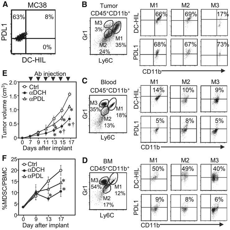
Infusion of anti-DC-HIL mAb retarded MC38 tumor growth and reduced frequency of DC-HIL+ MDSCs in tumor. A, Expression of DC-HIL and PDL1 on MC38 cells was assayed by flow cytometry. B–D, Cells isolated from tumors (B), blood (C), or BM (D) of MC38 tumor (~1.5 cm)-bearing mice were Ab stained and gated for CD45+CD11b+ cells (to exclude tumor cells), which were sorted into M1 (Ly6ChiGr1hi), M2 (Ly6CloGr1lo), and M3 (Ly6CloGr1hi) subsets and examined by flow cytometry for DC-HIL or PDL1 expression. E and F, On day 6 post-subcutaneous implantation of MC38 cells, mice (n = 5) were given intraperitoneal injection of UTX103 anti-DC-HIL mAb (αDCH), anti-PDL1 mAb (αPDL), or control IgG (Ctrl) every 2 days for a total of 6 injections. Tumor volume was measured every 2 days (E), and percentage of CD11b+Gr1+ MDSCs in PBMCs of blood on indicated days was determined (F). *, P < 0.01 and †, P < 0.01 compared with Ctrl and aPDL, respectively.
