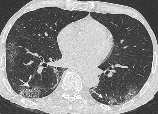
Fig. 9.

A 68-year-old man COVID-19 positive man presenting fever and dry cough for 7 days. CT scan shows multiple areas of panlobular GGO. In the posterior lower right lobe, an area of lung alteration with interlobular septal thickening resulting in a reticular pattern is present. Air bubble sign (arrows) is bilaterally recognizable. Air bubble sign consists of a small air-containing space, that can result from a dilatation of a physiological space or from lung cystic changes [24] or can be related to consolidation resorption
