Figure 1.
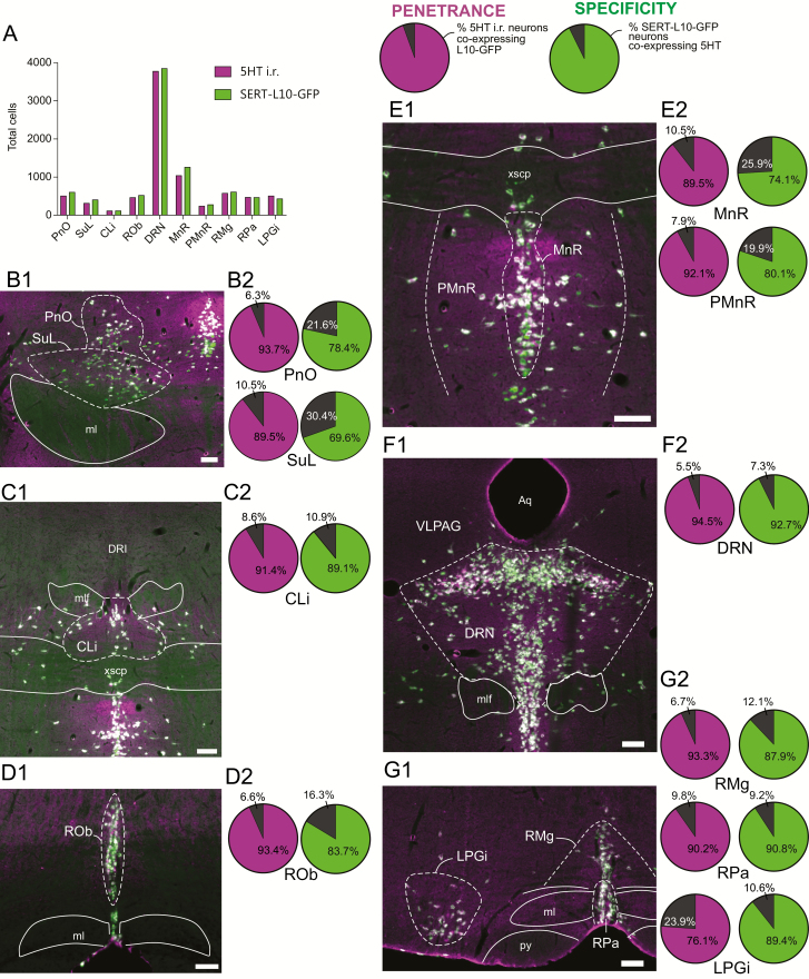
Validation of the SERT-cre mouse line. (A) Total number of 5HT immunoreactive (i.r.) neurons and SERT-L10-GFP neurons counted per brain region. (B–G) Left: Photomicrographs illustrating 5HT i.r. (magenta) and SERT-L10-GFP (green) in the pontine reticular nucleus, oral part (PnO) and supraleminiscal nucleus (SuL, B1), caudal linear nucleus (CLi, C1), raphe obscuris (ROb, D1), median raphe (MnR) and paramedian raphe (PMnR, E1), dorsal raphe nucleus (DRN, F1) and raphe magnus (RMg), raphe pallidus (RPa) and lateral paragiganticellular nucleus (LPGi, G1). Right: Pie charts depicting for each counted region the penetrance of Cre expression in 5HT i.r. neurons (i.e. the percent of 5HT i.r. neurons that expressed Cre-driven L10-GFP, purple shaded slice) and the specificity of Cre expression within each serotoninergic population (i.e. the percent of SERT-L10-GFP expressing neurons that were also 5HT i.r., green shaded slice). Scale bar: 100 µm. Abbreviations: Aq; aqueduct, ml; medial leminiscus, mlf; medial longitudinal fasciculus, py; pyramidal tract, xscp; decussation of the superior cerebellar peduncle.

