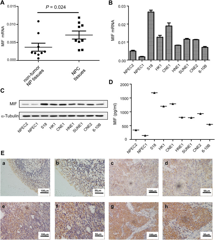
Figure 1.
Expression of MIF messenger RNA (mRNA) or protein in nasopharyngeal carcinoma (NPC) cell lines, and immunohistochemical staining of MIF in NPC tumor tissue. The levels of mRNA in non-tumor NP tissues and NPC tissues (A). The levels of mRNA and protein in the immortalized nasopharyngeal epithelial cell lines (NPEC1, NPEC2) and NPC cell lines were determined by real-time-polymerase chain reaction (PCR) (B), Western Blotting (C) and ELISA (D). Expression level was normalized by β-actin and α-tubulin, respectively. Error bars represent calculated from three parallel experiments. The normal nasopharyngeal epithelial tissue showed lower of MIF (E a-b). Low (E c-d), medium (E e-f), and high (E g-h) expression of MIF were showed in the nasopharyngeal carcinoma (NPC) tissues.

