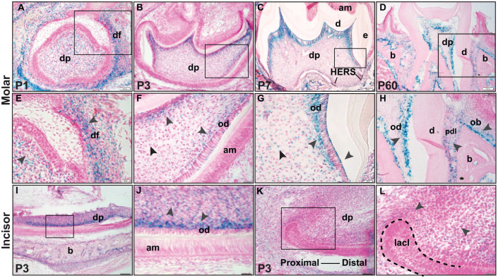
Figure 1.
Localization and expression of Ddr2 in dental and periodontal tissues. (A–H) X-gal staining of Ddr2+/LacZ mice reveals Ddr2 expression in the molars during development through adulthood. (A) At P1, Ddr2 expression is observed in dental papilla (dp) and the surrounding dental follicle (df) in developing molars (high magnification in E). (B–D) Ddr2-LacZ expression is selectively high in dentin-producing odontoblasts (od) at P3, P7, and P60. (F, G) High magnification also reveals Ddr2 expression in odontoblasts (od) and a subset of cells in dental pulp (dp). In a fully erupted molar (D, H), Ddr2 is expressed in PDL cells (pdl) and putative alveolar bone–associated osteoblasts (ob). Black arrowheads point to the expression sites. (I–L) Ddr2 expression in odontoblasts (od) of lower incisors, in dental pulp (dp), and around the labial cervical loop (lacl) at the proximal end of the incisor (dashed lines). (J, L) Higher magnification of boxed areas. No Ddr2 expression was seen in molar (F) or incisor (J) ameloblasts (am). Scale bar: 100 µm in A–D, I, K; 20 µm in E–H, J, L.

