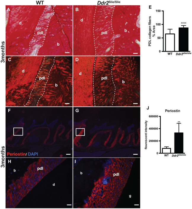
Figure 3.
Ddr2slie/slie teeth had atypical periodontal collagen fibers. (A–D) Picro Sirius Red (PSR) staining of periodontal ligament (PDL) collagen fibers in 3-mo-old mice under bright-field (A, B) and polarizing (C, D) microscopy. (E) Quantification of the PDL collagen area stained with PSR, n = 3 mice, 6 measurements per mouse. (F–I) Immunostaining of periostin (red) with inserts (H, I) showing periostin expression in the PDL space surrounding the molar roots. The cell nuclei were stained with DAPI (blue). (J) Quantification of the fluorescent intensity of periostin immunostaining, n = 3 mice, 5 measurements per mouse. **P < 0.01. ****P < 0.000. Scale bar: 200 µm in F, G; 20 µm in A–D, H, I.

