Figure 5.
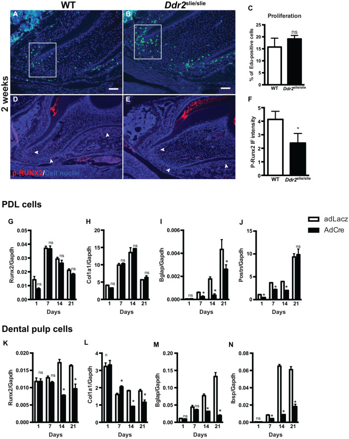
Ddr2 knockout exhibits decreased osteogenic and odontogenic differentiation of periodontal ligament (PDL) and dental pulp cells, respectively. (A, B) Representative images of EdU staining for labeling proliferating cells (green) and DAPI staining for cell nuclei (blue). (C) Quantification of the percentage of EdU-positive cells. (D, E) Representative images of p-RUNX2 immunostaining (red) in the nuclei of PDL cells and at the apex of the developing root. (F) Quantification of the fluorescent intensity of p-RUNX2 immunostaining, n = 3 mice. (G–J) Quantification of differentiation markers for PDL cells. (K–N) Quantification of differentiation markers for dental pulp stromal cells after 21 d. *P < 0.05. ns, not significant. n = 3 cell cultures/group. Scale bar: 50 µm in A, B; 100 µm in D, E.

