
FIGURE 1.
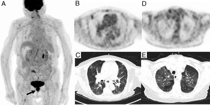
PET/CT acquisition was performed 1 hour after intravenous infusion of 145 MBq of 18F-FDG. A, 18F-FDG PET/CT maximum intensity projection showed an intense uptake (SUVmax 12.8) in anal canal primary neoplasm (black arrow) and increasing uptake in right lung corresponding in axial PET/CT slices to subpleural ground-glass opacity with 18F-FDG increasing uptake in right upper lobe (SUVmax of 2.4) (B, C), associated with patchy, rounded, diffuse, ground-glass opacity with mild FDG uptake (D, E). This incidental lung lesion finding, even in an asymptomatic patient, was consistent with typical appearance of COVID-19 infection during the March 2020 pandemic.1–5 There was no evidence of lymph node FDG uptake compared with some prior reported cases.6,7 The infection was confirmed by reverse transcription–polymerase chain reaction on nasopharyngeal swab collection.