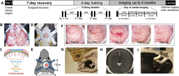
Fig. 1.
A) Experimental timeline. Each animal receives a chronic cranial window and head plate installation on Day 0, followed by 7 days of recovery and at least 4 days of training. On each day of the training, the animal receives two 1-hr training sessions composed of 3 steps to acclimate to head restraining. The animal returns to the cage to rest between training sessions. On the day of awake imaging, the animal receives an additional training before being head-restrained. The maximum time required for IOSI (with whisker stimulation), OMAG, DOMAG and Velocimetry (with whisker stimulation) is 15 min, 2 min, 5 min, and 15 min, respectively. The chronic cranial window can be imaged for several months. On the last day, the animal is anesthetized again, optional imaging can be performed. B) Microscope image of a newly installed cranial window. C) Cross-sectional anatomy of the craniotomy. D) Microscope image of a newly installed head plate. E) Schematics and dimension of the cranial implants. F) Series of microscope images of the cranial window from Day 0 to Day 14. G), H), and I) The 3-step handling/training to the head-restraining device. See Visualization 1 (65.4MB, mp4) for movie.

